


This website uses cookies to help improve your user experience


Machine learning, big data, and artificial intelligence aren’t just buzzwords in healthcare anymore, they’re becoming a core part of AI in healthcare operations. Hospitals and care teams are using these technologies to make sense of patient data, support diagnoses, optimize resources, and deliver consistent, high-quality care. What began as experimentation has now evolved into practical, regulated systems that are driving real improvements in patient outcomes and hospital efficiency.
Convolutional neural networks, support vector machines, and discriminant analysis algorithms empower doctors to predict and cure even the deadliest diseases. Apart from that, machine learning algorithms in healthcare help optimize other medical activities like administrative workflows, remote patient consulting (so-called telehealth), and healthcare data structurization.
In fact, Accenture’s Healthcare AI Adoption Insight report shows that 83 % of U.S. healthcare C‑suite executives are already testing generative AI in pre-production environments to boost productivity and smooth out clinical workflows1. It seems even hospital boards don’t want to wait for AI to finish its coffee before it starts working. It is a clear sign that healthcare is moving beyond experimentation, with AI becoming a strategic part of day-to-day operations.

The promise of Gen AI
Let’s explore four examples of machine learning in healthcare that are already improving or outperforming traditional human workflows.
Medical image analysis remains one of the most effective ways to detect cancer, strokes, lung disease, and other serious health conditions. Today’s machine learning and deep learning models can interpret CT, MRI, X‑ray, and ultrasound images with accuracy that often matches or surpasses expert human review.
Yet even with these advances, access to imaging remains deeply unequal around the world. Recent evidence suggests that roughly two‑thirds of the global population still lack reliable access to basic diagnostic imaging, especially in low- and middle-income countries where equipment and trained specialists are scarce2.
In some regions, there’s less than one CT scanner per million people, compared with around 40 per million in wealthier nations. In other words, some patients would have to fight a line longer than a Black Friday sale just to get a scan. This diagnostic divide means that life-saving imaging, and the AI tools that can enhance it, are simply unavailable to many who need them most.
Even where imaging exists, manually reviewing thousands of high‑resolution scans is a daunting task. Finding a subtle sign of disease among hundreds of slices on a CT or MRI still demands intense focus and experience. Machine learning helps by flagging suspicious features and patterns, allowing radiologists and pathologists to spend their time where human judgment matters most.
Studies show that deep learning algorithms are quickly reaching the level of human experts in medical image analysis. Trained on vast datasets, these models now achieve diagnostic accuracy above 90% for tasks like distinguishing malignant from benign tissue and classifying cancer subtypes3. In some breast cancer studies, AI systems perform on par with leading pathologists, demonstrating their ability to support crucial diagnostic decisions.
Unlike humans, neural networks are able to quickly learn on enormous image datasets. For example, IBM acquired more than 30 billion medical images to train its custom IBM Watson technology for early cancer identification.
Today, the most effective implementations pair deep learning tools with human expertise. In clinical settings where AI models act as “second readers”, radiologists and pathologists can focus on complex cases while AI handles the bulk of initial analysis.
Beyond accuracy, medical image analysis systems are also very cost-effective compared to a team of highly professional radiologists. AI doesn’t replace skilled clinicians, it helps expand access to reliable screening and interpretation, especially in places where expert care is hard to come by.
Let’s have a look at these two cases, proving that machine learning algorithms have already outstripped human doctors on their healthcare field.
Mentioning other AI use cases in healthcare, robot-assisted surgery is emerging as one of the most promising AI-powered technologies. Precedence Research analysts predict that the global surgical robotics market is set to grow, potentially reaching $50-66 billion by 20356.

Surgical robotics market size (2027 to 2035)
Surgeons have been relying on robotic systems for complex procedures for years, and their use keeps expanding around the world. The da Vinci® Surgical System, the most widely used platform, is now installed in over 7,500 hospitals globally, with millions of surgeries performed so far. These robots help make minimally invasive operations more precise, reducing complications, blood loss, and recovery time, so patients can get back on their feet faster than with traditional open surgery.
But what picture pops in your head when you hear “robotic surgery system”? Maybe you think of a robot operating a patient autonomously, while human surgeons play Mahjong and occasionally check things on the monitor. Unfortunately, this type of automation is yet to be achieved. A powerful surgeon’s mechanical arm, the da Vinci Surgical System is still not a fully-fledged medical robot.
As researchers say, a fully autonomous surgery system will require additional crucial steps. To become at least semi-autonomous, it must leverage the power of Big Data, ML/DL models, and implementation of computer vision. And that’s what AI engineers and data scientists have been working on for a very long time.
The concept of supervised autonomy, where human surgeons delegate certain tasks to a robot while overseeing its work, was first fully demonstrated with the development of the Smart Tissue Autonomous Robot (STAR). STAR, a semi-autonomous machine-learning-powered surgical system, became a benchmark in labs and experimental operating rooms worldwide. But can machine learning healthcare operations actually offer advantages over highly skilled human surgeons?
In 2025, STAR-derived autonomous robots performed complex gallbladder removals on realistic human-like models, completing all 17 procedural steps with 100% success across eight trials, adapting in real time to anatomical variations. Other studies have demonstrated millimeter-level precision in semi-autonomous tissue dissections and tumor resections, consistently matching or surpassing human performance. These results highlight that while STAR doesn’t replace surgeons, it extends their reach, enhances precision, and improves outcomes in highly complex procedures.
STAR is still not autonomous — it needs a tumor to be marked with the infrared markers. But STAR’s inventors promise that in just a few years robotic surgery systems will be extracting all the information they need from CT, MRI, and other medical images. It’s obvious that robots cannot replace human surgeons entirely in the nearest future. But very soon they will transform from a “mechanical hand” into fully-fledged surgeon assistants.

Transform AI concepts into production-ready healthcare systems. Oxagile designs and implements solutions that support clinicians and optimize operations.
We live in the age of sophisticated healthcare devices, from patient monitors and smart wearables to CV-powered scanners and internal biomonitors. Every moment, these devices generate a continuous stream of patient data including vital signs, activity patterns, and sleep. For years, much of this information went unused because traditional software could not keep up with the volume and complexity. AI and machine learning are now transforming this raw data into actionable insights that can make a real difference for patients.
AI-powered monitoring can detect subtle changes in a patient’s physiology, signaling early signs of complications or acute events such as arrhythmias or decompensation. Such early warning allows clinicians to intervene sooner, potentially preventing hospitalization or serious health crises. Studies in JMIR mHealth demonstrate that AI-powered wearables improve the prediction and detection of cardiovascular events and other conditions, giving patients and healthcare providers meaningful, real-time insights beyond the clinic walls7.
Machine learning, cloud computing, and big data are the three keystones of advanced patient data analytics in healthcare. Virtually unlimited resources of cloud computing support the effective storage and continuous processing of terabytes of patient data generated by remote monitoring devices.
Big Data algorithms structure and unify data. Then, machine learning takes the stage: it filters out data to generate important insights in real-time and automatically sends the most relevant information to the doctor. Moreover, deep learning applications in healthcare help facilitate treatment personalization by monitoring a patient’s health and carefully calibrating medical dosages when necessary.
Let’s witness another round of the Human vs. Machine battle to understand how machine learning solutions in healthcare help optimize 24/7 patient monitoring in the intensive care unit (ICU), the most important part of the hospital.
The intensive care unit is the place where human lives heavily depend on real-time data analysis. Today all the equipment in ICUs works separately, data sources and flows are not integrated, meaning all the equipment calibrations and adjustments must be made manually. Therefore, an effective treatment demands the ICU team’s undivided attention, but they simply cannot keep watch on every patient around-the-clock.
Innovators like Autonomous Healthcare are showing how AI and machine learning can pull together data from multiple monitors and clinical systems, giving caregivers a clear, real-time picture of a patient’s condition. Their tools (think AI-assisted ICU helpers) provide guidance on ventilation, fluid management, and hemodynamic support, helping clinicians keep patients in tip-top shape without breaking a sweat.
Researchers are even exploring AI-assisted drug dosing, though for now humans still have the final say. Even so, these systems already take a big chunk of the cognitive load off doctors’ shoulders, offering insights in real time that make critical care a bit less like juggling flaming scalpels.
Lots of doctors and nurses still handwrite chart notes and prescriptions, clinical notes and medical journals, and then have to transcribe the handwriting into a machine-readable format. This might as well be one of the machine learning healthcare use cases that can automate such processes to dramatically improve the effectiveness of clinicians, optimize their workflow, and reduce the costs of patient care.
Virtual personal healthcare assistants (VPHA) and telehealth solutions may be an answer. The standard workflow for virtual assistants is the following:
AI-powered virtual nursing assistants are able to give patients round-the-clock access to medical support and health monitoring.
Another way to optimize administrative work in hospitals is via the implementation of NLP algorithms. Voice-to-text and handwriting-to-text healthcare mobile apps are able to automate non-patient care activities like writing chart notes, prescribing medication, and ordering tests.
Doctors and nurses use NLP-powered healthcare apps to transform all types of clinical notes into machine-readable data for later ML analysis. Such healthcare automation with AI allows enriching actionable healthcare data and provides medical specialists with unprecedented timesaving capabilities.
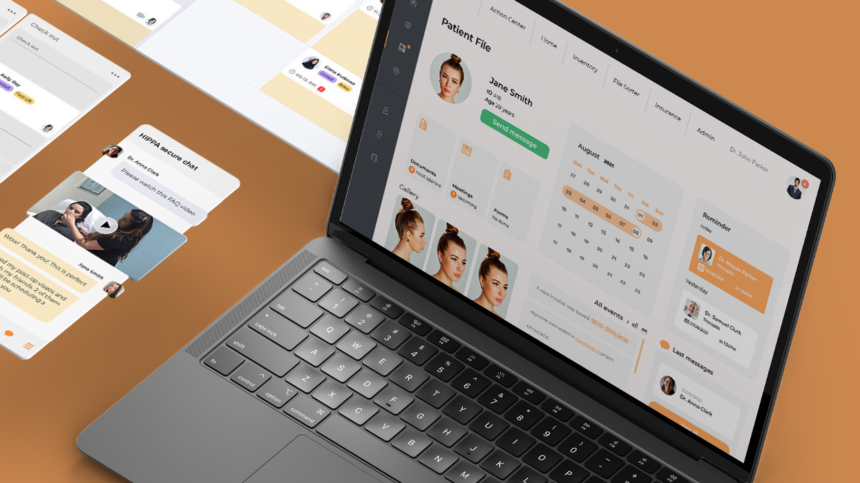
A fast-growing SaaS platform for aesthetic surgery clinics hit a scaling ceiling. Feature delivery slowed and clinics demanded smoother workflows and better patient experiences.
Our solution and impact:
Result: The team delivered a fully enhanced multi-device practice management system and outperformed the previous squad by 300%, accelerating feature delivery, easing administrative work, and improving patient satisfaction.
Computers are not about to replace doctors. Healthcare still relies on human judgment, intuition, and experience to interpret complex data. Physicians can observe subtle cues that machines cannot easily measure, such as patient behavior, body language, or even the smell of breath, all of which can inform critical decisions.
At the same time, we cannot underestimate the transformative power of machine learning in healthcare. AI is already delivering value in areas like medical image analysis, continuous patient monitoring, and administrative workflow optimization.
Some time ago, even the most advanced medical devices served primarily as tools for human operators. Today, AI, big data, and machine learning are shifting the paradigm, augmenting human expertise and providing more precise, efficient, and personalized care. Looking ahead, it is clear that already in the next five years, clinicians will increasingly rely on AI-powered assistants to handle major healthcare operations, not replacing them but extending their reach and impact.
Oxagile integrates AI in healthcare operations to streamline clinical and administrative workflows. Our solutions, including smart imaging, predictive patient monitoring, and automated records management, help hospitals deliver faster, safer, and more personalized care.
1. Gen AI amplified: Scaling productivity for healthcare providers — Accenture
2. Global radiology: Building equitable and effective partnerships with low and middle-income countries — Clinical Imaging
3. Deep Learning for the Early Detection of Invasive Ductal Carcinoma — JMIR Publications
4. Microsoft’s AI Is Better Than Doctors at Diagnosing Disease — Time
5. AI-Driven Real-Time Monitoring of Cardiovascular Conditions With Wearable Devices: Scoping Review — JMIR Publications
6. Surgical Robotics Market Size, Share and Trends 2026 to 2035 — Precedence Research
7. A systematic review and meta-analysis of artificial intelligence versus clinicians for skin cancer diagnosis — Nature

This is achieved by AI algorithms used to analyze patient data, forecast admissions, and optimize staffing and scheduling. By identifying potential bottlenecks using machine learning healthcare operations and reallocating resources in real time, hospitals can reduce wait times, improve bed utilization, and allow clinicians to focus on providing quality care.
Healthcare automation with AI reduces administrative burdens, minimizes human error, and allows staff to focus on patient care. With tasks like automated documentation scheduling, intelligent triage, and virtual nursing, AI-powered systems increase efficiency, lower operational costs, and free clinicians to spend more time with patients.
The list includes medical image analysis, predictive patient monitoring, personalized treatment recommendations, drug discovery, administrative workflow optimization, and AI in healthcare operations. These applications not only improve diagnostic accuracy and treatment outcomes but also boost efficiency and patient safety across the healthcare system.

