

Each medical imaging project draws on a set of established imaging techniques, selected against the diagnostic goal and the data available.
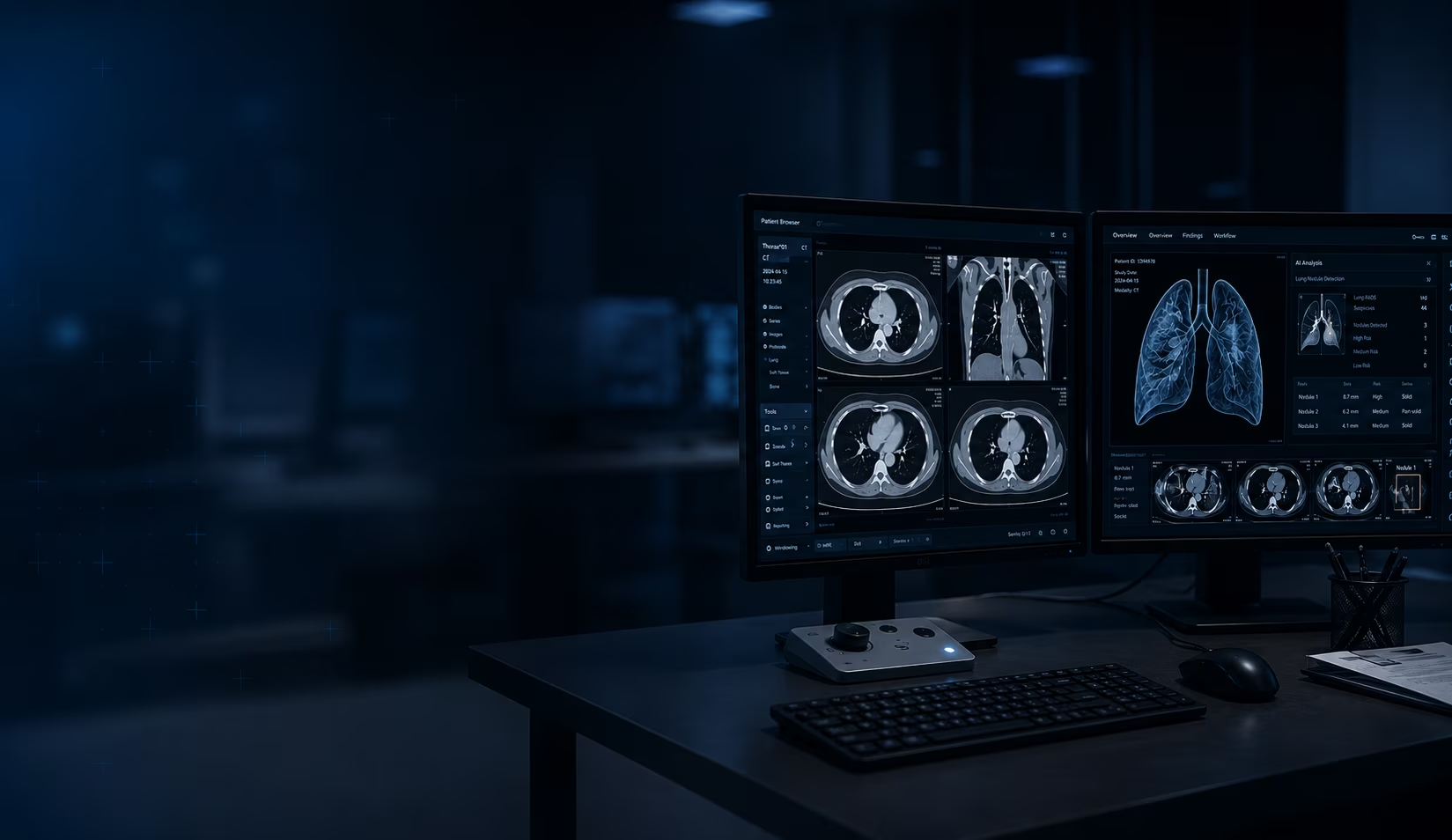
Different scanners, modalities, and capture conditions produce images that vary in character even when each is technically correct. A CT from one manufacturer, an MRI from another, a study captured under different contrast or positioning protocols, they all carry their own signal profile. Preprocessing normalizes these differences so the downstream analysis runs on consistent, comparable input regardless of where or how the image was acquired.
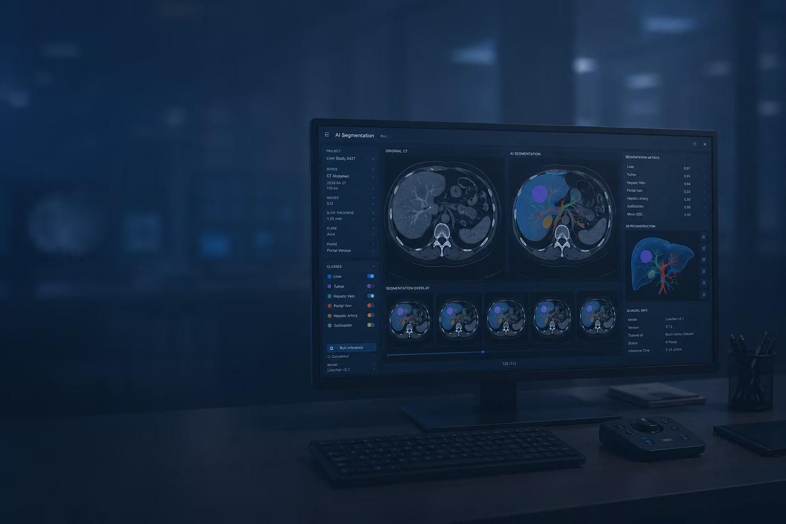
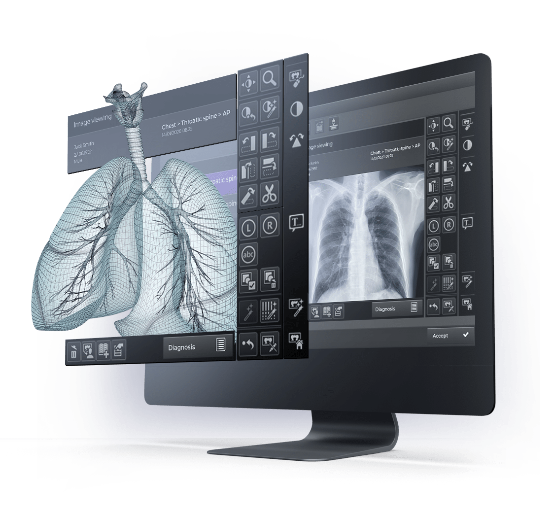
AI medical image segmentation separates a target structure, a bone, vessel, organ, or lesion, from the surrounding anatomy. Accurate segmentation is the foundation for quantification and any measurement a clinician acts on.
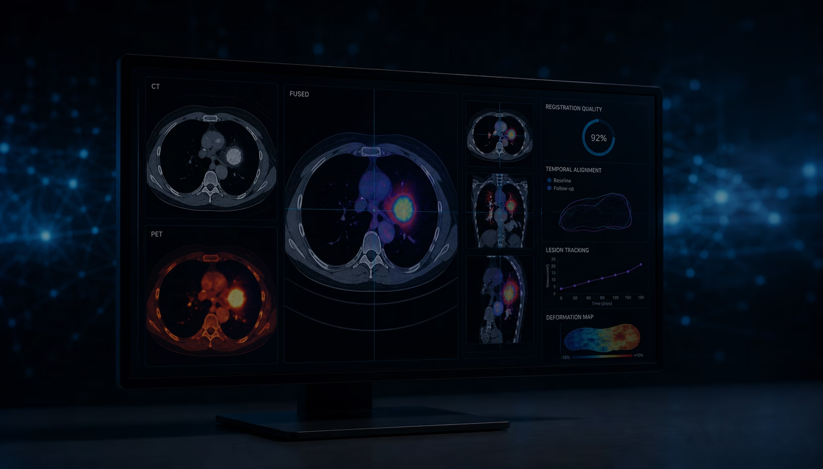
Fusion combines studies taken in different modalities, such as CT and PET, or the same modality captured at different times. Registration aligns these images to a common frame for tracking across a treatment course.
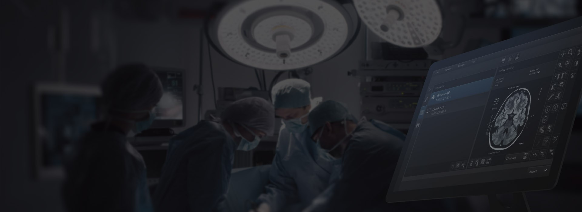
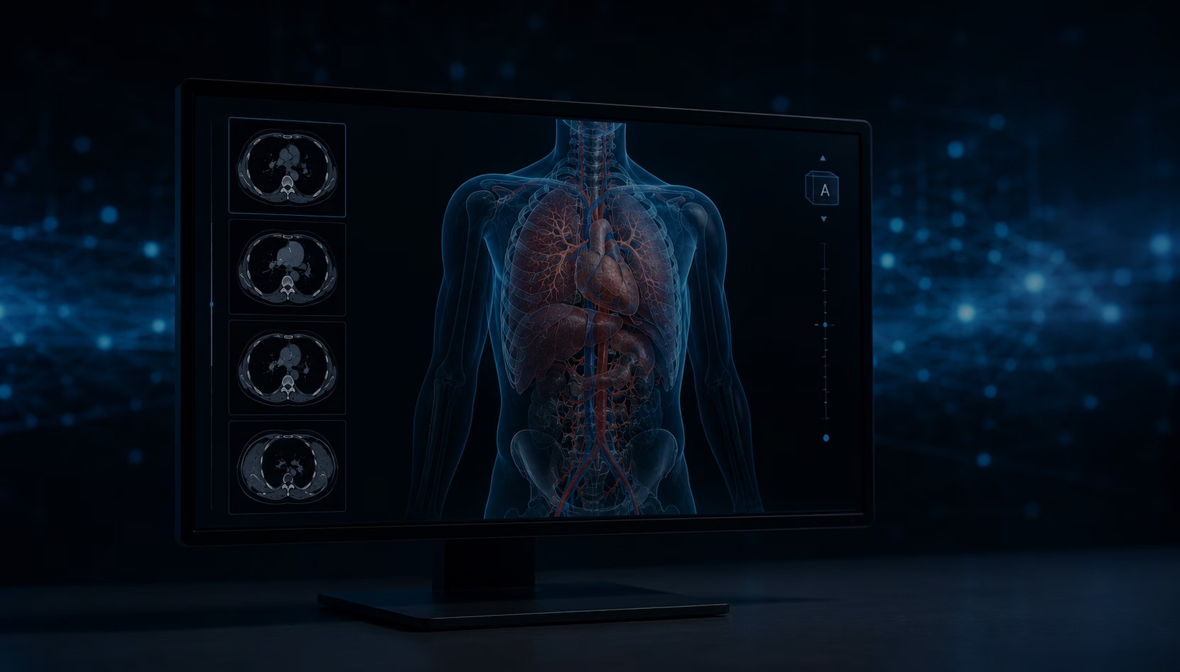
Oxagile converts CT and MRI series into high-resolution 3D models that can be examined layer by layer. Paired with AR and VR, these models support procedure rehearsal and intraoperative guidance, drawing on our computer vision and AR/VR practice.
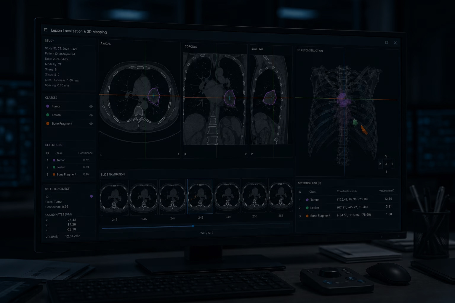
Objects of a defined class, a tumor, a lesion, a bone fragment, get located and their coordinates fixed, including position within a 3D model built from 2D slices. Quantification then characterizes each segmented structure and turns a visual read into measurable, repeatable values.
Discovery and clinical assessment
We map clinical goals, existing workflows, available datasets, and the regulatory requirements that apply.
Solution design and validation
Architecture, strategy, integration points, and success metrics are defined and pressure-tested.
Development and model training
Image analysis components are built and customized using the approach that fits the clinical problem.
Integration and compliance
The solution connects to PACS, RIS, and EHR/EMR systems through DICOM and standard interfaces with HIPAA alignment.
Deployment and ongoing optimization
Launch is followed by continuous monitoring, support, and model improvement as clinical needs shift.
Most imaging builds begin with a discovery call to map clinical goals, available data, and the systems involved. Oxagile’s experts can help clear things up.

As a medical image analysis software development company, Oxagile shapes every imaging project around clinical workflows from the first sprint. That’s what keeps it working past the demo and into a production setting.
Apply medical image analysis across specialties where early, accurate reads change patient paths.
ONCOLOGY Models trained on pathology images identify tumor types and mark suspicious tissue areas for review. The same analytical layer extends to laboratory studies, supporting immunohistochemistry and cell-level mutation analysis alongside the visual read.
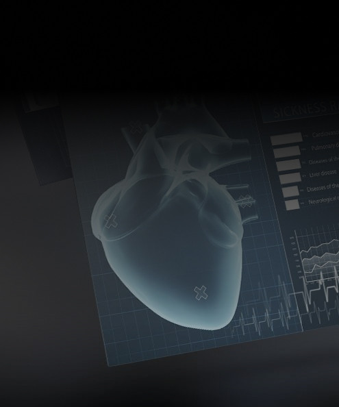
CARDIOLOGY Coronary artery segmentation supports early detection of vessel narrowing and hardening, giving cardiologists a window for intervention before a cardiac event.
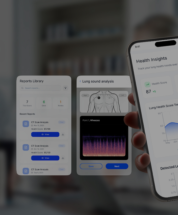
PULMONOLOGY Two- and three-dimensional processing of the lung capillary system supports detection of respiratory conditions such as emphysema and pulmonary embolism. Tracked over time, it also flags early signs of disease progression and acute exacerbations before symptoms escalate.
ORTHOPEDICS 3D bone alignment supports fracture surgery planning and provides digital guidance during the procedure.
OPHTHALMOLOGY Analysis identifies retinopathy of prematurity, retinal vein occlusion, anterior segment disorders, and other eye conditions, and supports computer-vision-assisted cataract surgery.
DICOM • NIfTI • Analyze • MINC • ECAT7
TensorFlow • PyTorch • MONAI • Keras
OpenCV • ITK • scikit-image • 3D Slicer
AWS • Azure • Google Cloud
HL7 • FHIR • HIPAA • IEC 62304

Built well, imaging software earns its place in the healthcare processes. Oxagile's work focuses on the gains that reach the provider and the patient: faster reads, fewer missed findings, and results inside the systems already in use.
Automated analysis takes over repetitive review tasks and shortens the time between scan and read. Clinicians spend their attention on the studies that need judgment.
Compliance and security are designed in from the first sprint, aligned with HIPAA, IEC 62304, and the standards that govern software in clinical use.
Consistent, reproducible analysis gives clinicians a second read that does not tire or vary by shift. Subtle patterns the eye can miss get flagged for review.
Imaging software earns its place by fitting the stack around it. Oxagile builds for integration with PACS, RIS, and EHR/EMR systems so results land where clinicians already work.

Compliance is built into design, not bolted on after. That covers encryption of data at rest and in transit, role-based access control, audit logging, and data handling aligned with HIPAA and, where relevant, IEC 62304 for software in clinical use. Documentation is produced alongside the build so audits do not stall delivery.

Yes. Oxagile builds integration layers that connect custom models to PACS through DICOM and standard interfaces, so inference results return to the same viewer and worklist already in use. RIS and EHR/EMR connections follow the same approach.

Yes. For surgical navigation and other latency-sensitive cases, models can run at the edge, close to the imaging hardware, so guidance keeps pace with the procedure. Model size, hardware, and accuracy targets are set against the clinical requirements during design.